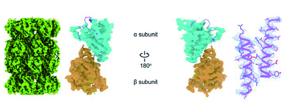
Cryo-electron microscopy (Cryo-EM) is an important tool to understand biological mechanisms at molecular level. However, cryo-specimen preparation still remains many challenges. Biomolecules tend to be adsorbed to the air-water interface when applied onto canonical supporting films in cryo-EM, causing partial denaturation and/or preferential orientations. In this study, we developd a new type of cryo-EM supporting material using bioactive-ligand functionalized single-crystalline monolayer graphene membranes as supporting films. We have demonstrated that the functionalized graphene membrane (FGM) has the superior property of high conductivity and mechanical strength when irradiated in electron microscopy, and high affinity and selectivity on target biomolecules. These merits effectively facilitate to prevent biomolecules from touching air-water interface, and enable us to get a near-atomic resolution 3D reconstruction. We anticipate that the FGM grids could make it more reproducible and robust to perform cryo-EM specimens, therefore enhancing the efficiency and throughput of high-resolution cryo-EM structural determination.
External Links
Internal Links
-
News
- Cao et al. Structural basis of endo-siRNA processing by Drosophila Dicer-2 and Loqs-PD, Nucleic Acids Research, 2025
- Zheng et al. Cryo-electron tomography reconstructs polymer in liquid film for fab-compatible lithography, Nature Communications, 2025
- Zheng et al. Self-assembled superstructure alleviates air-water interface effect in cryo-EM, Nature Communications, 2024
- Yang et al. Electrospray-assisted cryo-EM sample preparation to mitigate interfacial effects, Nature Methods, 2024
- Xu et al. Graphene sandwich-based biological specimen preparation for cryo-EM analysis, PNAS, 2024


